We are seeking volunteers comprising 40 ME/CFS patients and 20 controls to participate in an NIH funded (U54AI178855) project. An overview of the study can be found here. Only those participants whose onset of ME/CFS was before 2020 will be eligible. Please review our recruitment flyer for more information on how to volunteer as a patient or control in this research study.
Out in the Journal of Translational Medicine, Betsy Keller et al. describes results from the Center’s large multi-site 2-day CPET study. The publication analyzes cardiopulmonary exercise testing (CPET) data collected over two days from 84 individuals with ME/CFS and 71 sedentary controls. Within this population, 55 sex, age, and fitness-matched pairs were also compared. This is the largest 2-day CPET study of ME/CFS to date.
The article provides robust documentation of impaired recovery in people with ME/CFS following exertion (i.e., post-exertional malaise), something that is not seen in the control population. The study also recruited sedentary controls and used sex, age, and fitness-matched pairs to validate that fitness level does not predispose someone with ME/CFS to exertion intolerance. Keller et al. highlights that the autonomic nervous system is, at least partly, associated with ME/CFS due to the disrupted hemodynamic and ventilatory responses to exertion. Additionally, the worsening of CPET parameters over the two-day protocol led to increased clinical impairment status in ME/CFS whereas controls remain relatively the same.
Overall, the manuscript provides a detailed look at the physiological component of post-exertional malaise in ME/CFS. The paper is open access. Individuals interested in the topic should check out the full publication for more information.
Maureen Hanson spoke at the Demystifying Long COVID North American Conference, sponsored by Academic Medical Education organization, and her talk comparing ME/CFS with long COVID is now available online.
Center investigators presented at the 1st International Conference on Clinical and Scientific Advances in ME/CFS and Long COVID. Details regarding the agenda and event can be found on the event website. The two-day hybrid event was recorded. Both days of the conference are available below.
Day 1 (April 3, 2024): Dr. Susan Levine presented “Brief History of ‘Pandemics’ Related to ME/CFS Punta Gorda Florida and Royal Free Hospital in London. Origin of Current Case Definition.” Her talk starts at 27:00. Dr. Levine also took part in a panel discussion and “Conversation: The Role of Physician as Advocate, Lessons Learned from 30 Years of Clinical Practice” that occurred later in the day.
Day 2 (April 4, 2024): Dr. Maureen Hanson presented “Circulating Signals of ME/CFS.” Her talk starts at 6:07:00.
Exertion intolerance and post-exertional malaise are defining features of ME/CFS. Work from our Center aims to uncover the molecular disruption that occurs during and around these features. Thus, through the lens of extracellular vesicles (EVs), a publication from co-lead authors Ludovic Giloteaux and Katherine Glass provides novel insights into these topics.
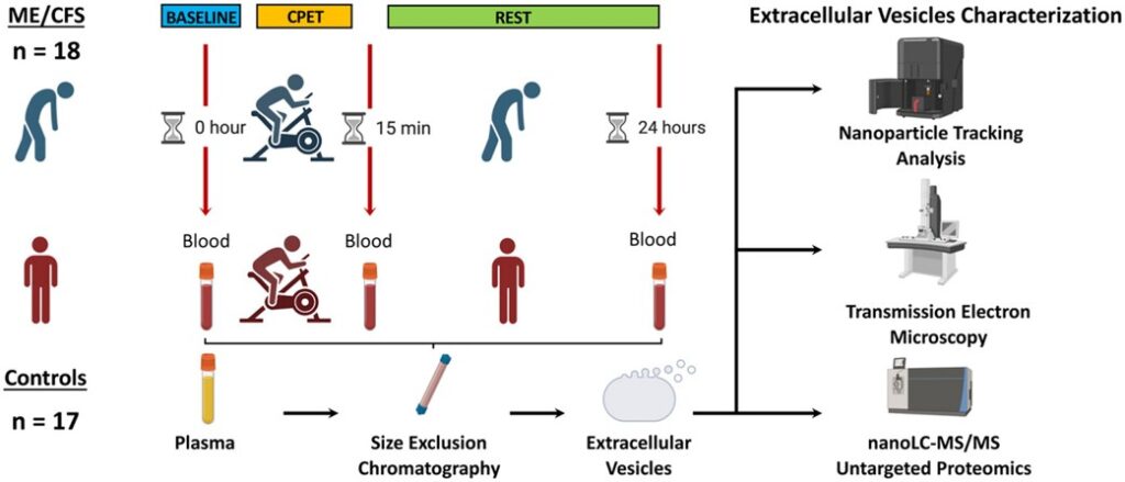
The study involved the isolation of EVs, membrane-bound non-replicating particles released from cells, from the plasma of 18 ME/CFS and 17 healthy control female participants. The cargo of EVs are relevant in that they provide signals that could uncover elements of dysfunction in ME/CFS. Specifically, protein cargo from EVs were examined in this publication before and after a cardiopulmonary exercise test (i.e., around the induction of PEM).
Figure 1 from the publication provides an overview of the study’s experimental design
The EV proteome in response to exercise was clearly different in ME/CFS when compared to sedentary controls. These divergent responses were associated with different molecular pathways. Uncovered protein differences between the cohorts also point to contrasting tissues and cell types. Additionally, there were several proteins associated with a worsening of muscle pain (e.g., THBS1 and TPM4), PEM (e.g., NEXN), and fatigue (e.g., CLU) post exercise in ME/CFS. Although it is difficult to directly associate changes with the EV proteome to distinct cell types, this publication brings forth avenues to interrogate further.
If you are interested in more information regarding this publication, the entire paper is freely available in the Journal of Extracellular Vesicles.
The working group of the National Advisory Neurological Disorders and Stroke Council, part of the National Institutes of Health, recommended the development of research priorities for ME/CFS to help move the field towards translational research and clinical trials. The result led to creation of the ME/CFS Research Roadmap Working Group. The Group, co-led by Drs. Lucinda Bateman and Maureen Hanson, produced a series of eight webinars. A complete description of the webinar series can be found on the ME/CFS Research Roadmap website.
Our Center’s mission is to promote research to identify its cause(s), biomarkers, and pathophysiology in order to lead to prevention and effective treatments. With this focus, several Center investigators presented data to support the development of research priorities for ME/CFS. The talks from our Center are included below.
Maureen Hanson presented “ Immune cell-type approaches to identify mechanisms of ME/CFS” during the Immune System webinar. Her talk starts at 1:38:40.
Jessica Maya presented “Investigations and Consequences of Altered Metabolism in ME/CFS Immune Cells” during the Metabolism webinar. Her talk starts at 58:32.
Maureen Hanson presented “Chronic infection in ME/CFS: non-Herpes viruses” during the Chronic Infections webinar. Her talk starts at 1:01:25.
Ludovic Giloteaux presented “Extracellular vesicles” during the Physiology webinar. His talk starts at 3:15:10.
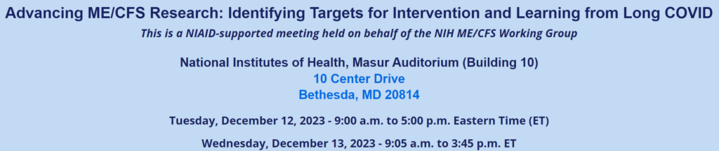
NIH held a hybrid online conference on the NIH campus on December 11-12, 2023. Center personnel attending in person were Claire McNally, Annie Gardella, David Iu, Jessica Maya, Tien Luyen (“Louis”) Vu, Katherine Glass, Arnaud Germain, Ludovic Giloteaux, Dawei Li, Andrew Grimson, and Maureen Hanson, as well as collaborator Nicholas Hampilos from Weill Cornell Medicine.
NIH sponsored an early career researchers workshop on December 10, 2023 attended by Cornell graduate students Claire McNally Annie Gardella, and David Iu, postdoctoral associates Jessica Maya and Tien Luyen (“Louis”) Vu, and Research Associates Katherine Glass, Arnaud Germain and Ludovic Giloteaux. Drs. Glass and Maya helped organize the meeting.
Verbal presentations:
David Iu: Epigenetic Reprogramming of CD8+ T cell Populations Drives Exhaustion in Myalgic Encephalomyelitis/Chronic Fatigue Syndrome (ME/CFS)
Tien Luyen Vu: Single-cell transcriptomics of ME/CFS circulating immune system before and after symptom provocation
Poster presentations:
Anne Gardella: Cell-free RNA signatures of myalgic encephalomyelitis/ chronic fatigue syndrome
Ludovic Giloteaux: Extracellular vesicle protein cargo in ME/CFS cases and controls following maximal exercise
Arnaud Germain: Proteomic adjustments following induction of post-exertional malaise
Claire McNally: Investigating the role of iNOS in endothelial dysfunction in ME/CFS
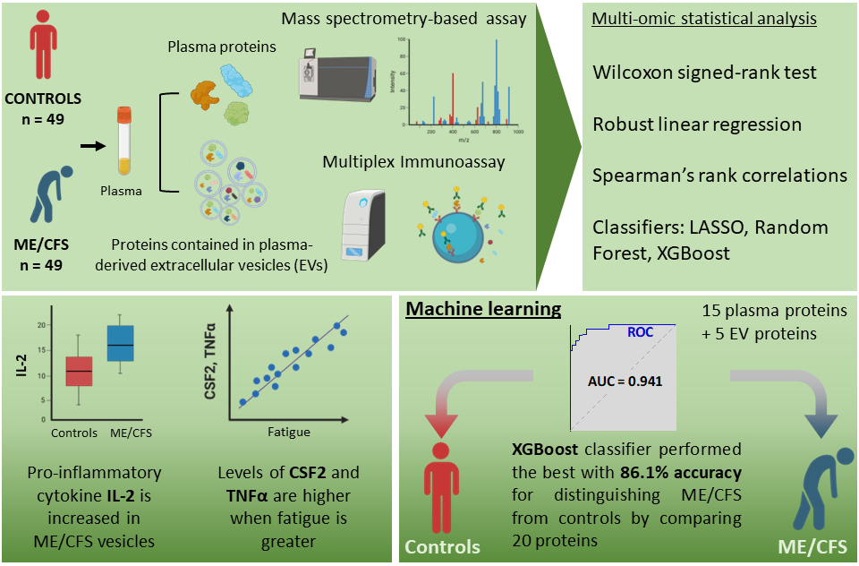
A new open access publication in the Journal of Translational Medicine describes the work by Giloteaux et al. to uncover ways to detect the disease ME/CFS. Ludovic Giloteaux and Jiayin Li, joint first authors, took a collaborative approach to improve our understanding of ME/CFS. Giloteaux isolated extracellular vesicles from the plasma of 98 Chronic Fatigue Initiative individuals (49 ME/CFS and 49 controls) to study their signaling molecules (i.e., cytokines). Then he worked with Jiayin Li and David Ruppert, statisticians at Cornell, and using data generated by Columbia University investigators, the group combined plasma cytokine, EV cytokine, plasma proteomic, and demographic datasets to explore new ways to approach ME/CFS.
Ludovic Giloteaux
One of the key findings from the publication is the 86% accuracy in differentiating between people with ME/CFS and health controls. Giloteaux et al. leveraged multiple datasets to achieve this goal. The paper also outlines interesting correlations between various biological molecules and clinical surveys that measure disease severity. For example, higher levels of pro-inflammatory molecules (e.g., CSF2 & TNFa) were correlated with greater physical and fatigue symptoms in people with ME/CFS.
The publication is open access so see the website for more information. Additionally, the EV cytokine data is available on mapMECFS.
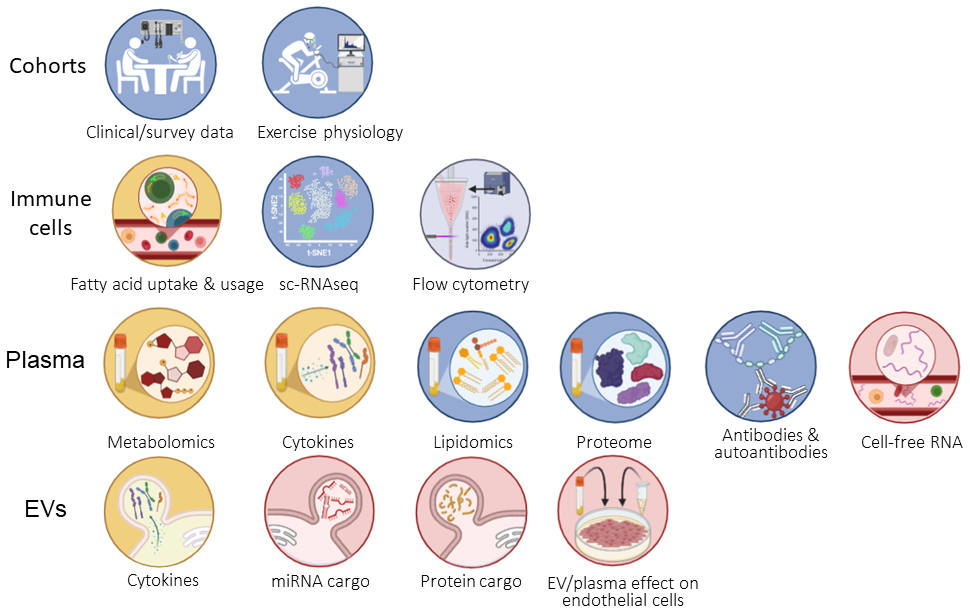
Announced April 11, 2023, the ENID Center has successfully competed for a 5-year U54 award from the National Institutes of Health. The U54 award provides funding for a multidisciplinary, multicomponent collaborative research center. The award will fund exciting research to explore topics such as endothelium function, cell-free RNA, immune cell dysfunction, extracellular vesicles, and more.
The new research award includes a subject participation component. We will soon provide information on how interested people can get involved. Check back here later, or stay tuned to the Center’s tweets and Facebook posts for updates.
The new funding is partly an extension of previous work. Specifically, we plan to utilize previous and future data, highlighted in the figure below, to perform multiomic analyses. Multiomics uses sophisticated computation approaches to incorporate multiple datasets, which can provide an enhanced and holistic perspective.
The Cornell Chronicle first announced the U54 award. Check out the press release for more information.