The discovery of ME/CFS-specific biomarkers that can advance our understanding of the illness and its pathogenic mechanisms, differentiate the disorder from overlapping or comorbid diagnoses, and identify potential treatment targets is currently a pressing and unmet research and public health need. We are working to fill this knowledge gap by using advanced neuroimaging techniques to test and then validate a pathophysiological model of ME/CFS which posits that oxidative stress and neuroinflammation are intertwined mechanisms in the etiopathogenesis of the disorder.
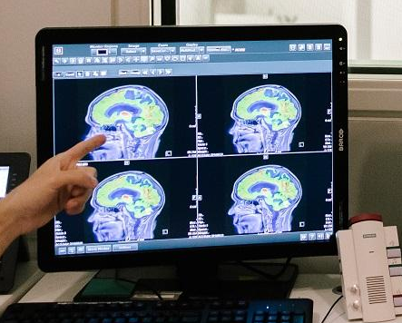 We have used proton magnetic resonance spectroscopy (1H MRS) to measure in vivo brain levels of glutathione (GSH) -- the most abundant antioxidant in the central nervous system – as a marker of oxidative stress. The same method was used to measure in vivo brain levels of lactate and N-acetylaspartate (NAA) as markers of mitochondrial dysfunction. To measure in vivo brain levels of ATP, creatine phosphate (PCr) and inorganic phosphate (Pi) as complementary indices of mitochondrial dysfunction, 31P MRS was used. In vivo brain 11C-(R)-PK11195 positron emission tomography (PET) was used to measure the binding potential of the ligand as a marker of neuroinflammation. We are examining the levels of circulating markers of neuroinflammation and oxidative stress for corroborating the proposed neuroimaging biomarkers.
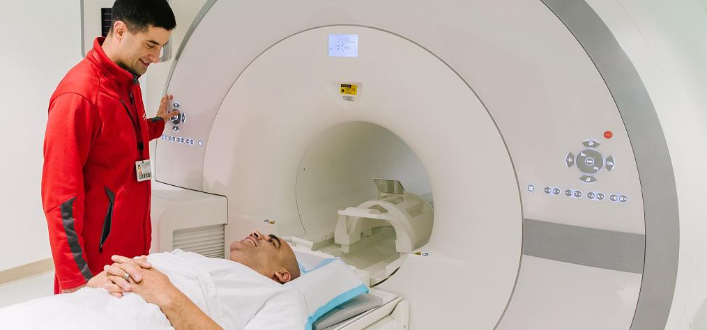 Our analyses was performed before and following symptom provocation with cardiopulmonary exercise tests (CPET), known to trigger post-exertional malaise (PEM) in ME/CFS patients. This research has the potential to shed new light onto the neurobiological mechanisms and underpinnings of ME/CFS to advance our understanding of the illness, and establish ME/CFS-specific biomarkers for differentiating the disorder from overlapping or comorbid diagnoses, and identifying potential treatment targets.
This NIH funded (U54NS105541) project is led by Dr. Dikoma Shungu, an accomplished magnetic resonance spectroscopist/physicist and computer programmer. In addition to prior work on ME/CFS, he has previously used neuroimaging methods to study fibromyalgia, Gulf War Illness, schizophrenia, major depression, anxiety and obsessive-compulsive disorders, Parkinson’s disease, ALS, primary mitochondrial disorders, and brain tumors. He is assisted by Xiangling Mao, a long-term associate and computer programmer skilled in analysis of spectroscopy data. Dr. Yi Wang, an internationally known theoretical physicist, is playing a major role in assessing brain tissue iron content by MRS.